Jun 5, 2014

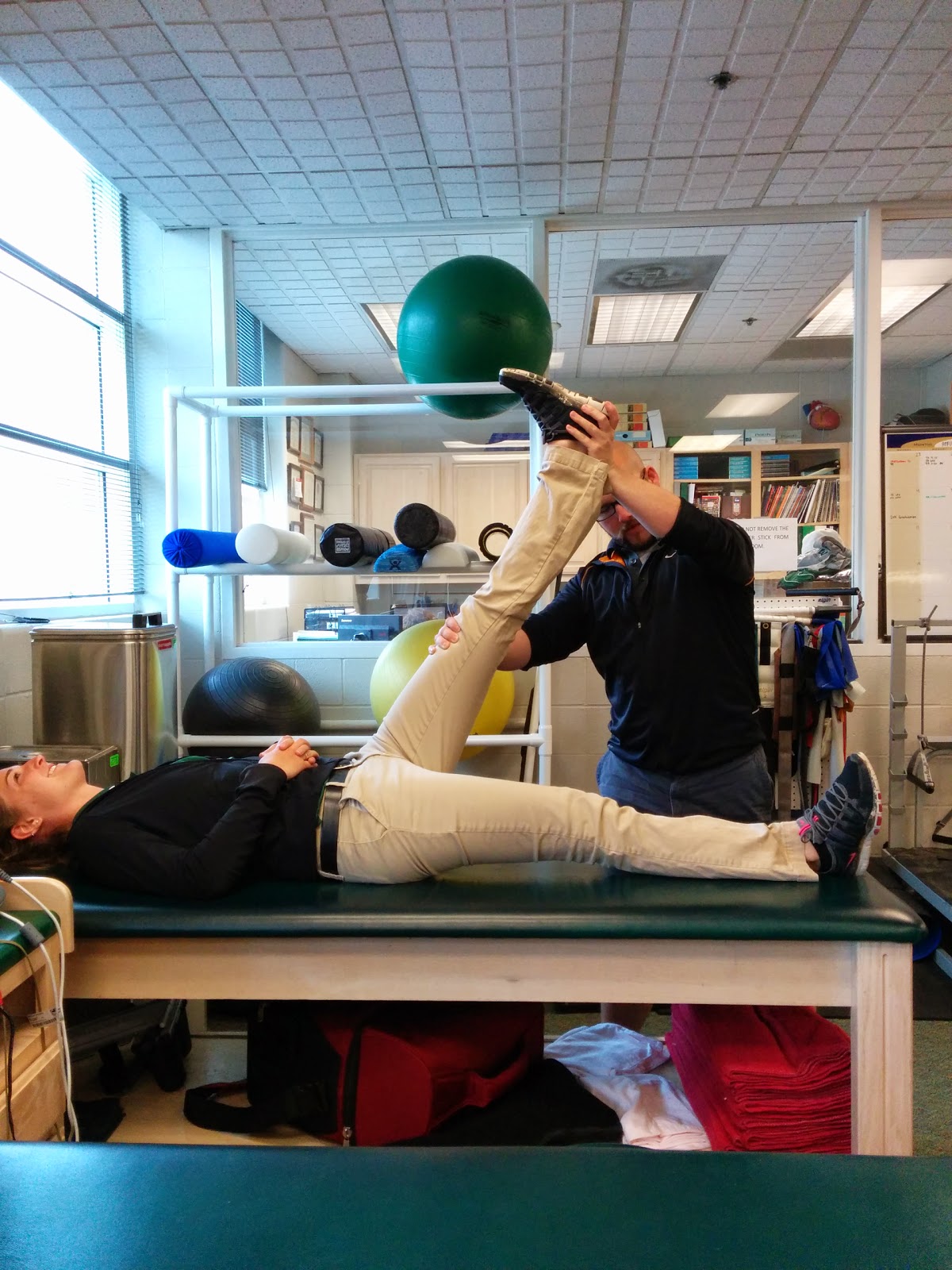
Tibial Internal Rotation Mobilization w/ The Edge Mobility Band


I have had a few people ask me lately about using the Edge Mobility Band for tibial internal rotation. I kept trying to refer them to a previous video that I had made about this very topic. However, they couldn't seem to find the video on my blog or on my YouTube channel. Turns out I never edited the video or posted about it on my blog. Oops.

Therefore, this is a quick post and video to show you how I use the Edge mobility band to work on mobility deficits when it comes to tibial IR. A few things before I share the video...


-The band is not a necessity for this technique but I find that it helps enhance its efficacy and the ability to grip the skin.
-The band doesn't need to be wrapped on so tightly that it cuts off blood flow.
-This technique should be pain free.
-You are looking to get at least 20 degrees of tibial internal rotation via the SFMA to be functional...Don't confuse a mobility issue with a motor control issue here. (Active vs. Passive differentiation)
-Do more than the two sets of ten that I did for this video. I filmed this quickly on my phone while at work tonight using one of my coworkers. In the first segment of the video he is actually trying to internally rotate his tibia. He definitely isn't functional afterwards but there is a marked improvement afterwards. Rinse and Repeat this continuously for a few days to weeks to restore full motion.


I am curious what others do for mobility issues regarding tibial IR and subtalar inversion/eversion issues...Please share if you have some input!

Jun 1, 2014

5 Tips For Improving Great Toe Mobility and Stability

The foot on the right is far from ideal, but its great toe position is better


In my previous post I discussed the importance of the "big", "1st", "great" toe, or hallux. I finished that post with a promise to share some ideas and methods for enhancing the health of your foot/toes and here I am to make due on that.

In reality, some people may already be too far gone to see much benefit from conservative methods of treatment. Unfortunately, with enough abuse from faulty footwear, poor biomechanics, and overuse some feet may require surgery. That doesn't mean that you may not find a decrease in symptoms or pain from conservative methods and all avenues to avoid surgical procedures should be explored in my opinion.

Instead of giving tons of in-depth and intricate details on things that you can do for better foot/toe mobility and motor control I am going to give some simple and easy ideas and concepts that can have a huge impact.

1) Avoid Shoes With a Tapered or Narrow Toe Box

The toes should be the widest part of the foot. This is often a rarity for people of developed nations and many think it is normal. However, if you look at a young child or toddler's foot it is likely that their foot still holds this true form. If you look at most footwear you will find that the toe boxes are often very constraining and the widest part of the shoe is usually not at the toes. They often come to a very distinct point about where the second toe is. One thing that I recommend when picking out a pair of shoes is to perform the "shoe liner" test. This is where you remove the liner or insole of the shoe and place your foot on top of it. If your toes splay over the edges then it is likely that the shoe is too narrow for your foot. This is very important for the developing feet of children and I think this video by Dr. Ray McClanahan does a great job of explaining this.

2) Avoid Shoes With a Toe Spring Design

If you view a shoe from a side profile you will notice that many conventional shoes will have a design where the sole will curve upward near the toes. This places the toes into an extended position relative to the metatarsals when at rest. I believe this was originally designed to help the shoe have a "rocker" effect to help people with poor ankle mobility.

3) Avoid Shoes With an Excessively Elevated Heel, "Ramp", or Heel-Toe Drop Design.

I attribute this shoe design and our cultures disdain for squatting (excessive sitting in chairs) to be major contributors to the poor ankle mobility that plagues many people and athletes alike. These three design flaws are discussed indepthly by Dr. Ray once again and he does a great job discussing them in this other video of his. You can do all the joint mobilizations, stretching, and flexibility work in the world but if you keep yourself or your patients in crappy shoes then you are fighting a losing battle.


4) Ensure Adequate Ankle Dorsiflexion

One thing that I have noticed in many patients with issues in the toes is that they are often accompanied by issues at the ankle, especially regarding decreased ankle dorsiflexion. Chicken or the egg here? I'm not sure but if a person lacks the ability to achieve full ankle dorsiflexion then their toes may be at risk for taking an extra beating. What about if you lack toe mobility as well? Good Luck. Here is a video I made for a quick and easy way to check and see if you have enough ankle dorsiflexion.


5) Increase foot intrinsic muscle strength/motor control

It isn't advisable to dive in headfirst and make drastic changes to your footwear in one fell swoop and expect all to be well. Even if you aren't going to change your footwear choices it is still probably advisable to work on intrinsic muscle strength/control. Here is another video I made previously showing some easy ways to improve foot/toe strength and mobility.


If you are still struggling with issues in your feet, ankles, or further up the kinetic chain and these simple tips aren't enough to resolve them then you may possibly need additional manual therapy from an athletic trainer, physical therapist, physiotherapist, podiatrist or chiropractor. There is also the possibility that a conventional approach may not be enough for some issues.

Some issues may require more time or help from products like Correct Toes. An foot-toe orthosis like them requires a post all to themselves but I personally have used it on myself, my patients and in research with some surprising success and outcomes. One thing that I will say is that I like how that product versus the cheaper generic types that you can find at say Walgreens or CVS will actually fit in your shoes, has multiple sizes, and can be modified for your foot shape.


May 20, 2014

How Healthy is Your Big Toe?

Hallux Valgus is only good for suppressing appetite and skinny pumps

Proper motion, strength, and position of your toes is a concept that is often lost with conventional approaches to health care. The toes may be one of the most commonly disfigured series of joints in the body, especially the big toe. Dr. Perry Nickelston of Stop Chasing Pain refers the big toe as one of the key movement linchpins in the body. For those of you that are unaware of linchpins it is the key piece of a complex system which is vital for holding it all together.

In reality, the above pictured toes should not be as common as they are. Right away the thoughts stray to women who indulge themselves with high heeled or skinny footwear with narrow and tapered toe boxes for fashion. During medieval times the fashion was for men to wear long, narrow and pointy shoes (think like elves?) and cadaveric studies of people from this time show that these disfigurements were more common in men than women which is the exact opposite of modern times/studies.

These sandals are a little too small for this fellow.

A foot or more specifically your toes should ideally look a lot more similar to the above foot. In fact, there really should be a slight gap between your 1st and 2nd toes! Having a big toe that is straight and mobile is beneficial for many different activities. In fact, I would argue that it is probably important for all activities that involve bearing weight through the feet. I've also seen videos of indigenous cultures using their toes with great dexterity.

"Sign here please, sir."
Your big toe is essential for proper balance, running, walking and many athletic activities or exercises. Somewhere around 55-60% of your support should come from your big toe during running. It also helps with foot stability, and explosive propulsion during power movements such as squats or the deadlift. Many bodyweight exercises or yoga positions require a mobile and correctly positioned big toe as well. You should have three major points of contact to the ground through your feet for proper function: the big toe, the calcaneus, and the lateral aspect of the foot around the base of the 5th metatarsal.

I dare you to try barefoot pushups without adequate toe mobility. Ouch.
I'm sure there are many people out there that are looking down at their feet and are taking notice of a slight to severe hallux valgus angle, or currently suffer from conditions such as hallux rigidus or limitus. You may even be functioning quite well in your prospective sports or activities of daily living. However, I can guarantee that a few things are probably true for you. 1) You are compensating for your poor positioning and/or mobility. 2) Your performance is lacking and could only be improved with improvements made here. 3) Finally, there is a chance you may be part of the 15%ish of the population that presents with Morton's foot.


If this is the case your 2nd toe will be longer than your first toe. In this circumstance your second toe is now taking an excessive load compared to what it was designed for. This is genetics at play and is arguably a less evolved toe structure from when the big toe was opposable like a thumb similar that of other primates.

In conclusion, the big toe can have a big impact on your health, performance and movement all the way up and down the kinetic chain. Even if you are not an athlete proper big toe health will be paramount when you reach an older age. The elderly are at risk for falling which has very serious impacts on life quality and expectancy post falling. Proper position of the big toe increases stability by widening the base of support and proper mobility of the big toe gives greater proprioceptive input for better neuromuscular control. Both are critical concepts for everybody.

In my next blog post/videos I will be going over ways to promote increased toe mobility and position! Stay tuned for more...

May 14, 2014

Thoracic Extension Doesn't = Thoracic Extension

Howdy Folks! Today I wanted to take a quick moment to comment on some discrepancies that I have noticed when hearing people discuss thoracic mobility and the need for thoracic extension. Anyone that is familiar with the Functional Movement System is probably well aware of the lack of thoracic mobility that many people seem to suffer from. This is something that you often see targeted by FMS corrective exercises or may be a common finding during an Selective Functional Movement Assessment (SFMA) for some.

On the other hand, there is the kind folks associated with the Postural Restoration Institute (PRI) that are trying to promote thoracic flexion and minimize hyperextension of the thoracic spine. Then I have heard stories from colleagues at PRI courses mention how they are lacking thoracic extension only to be told they have too much. So what is the answer? Do we need thoracic extension? Who is right here?

Well I believe that both of these systems or schools of thought are trying to achieve the same thing, and are essentially saying the same thing despite it sounding different. I am arguing that people are not differentiating between the different hinge points of the spine and the exact levels of the thoracic spine that they are referencing. Look at this first squat picture below. Nobody from either school is going to like this squat form and PRI'ists will notice the excessive thoracic extension from T8 and down while FMS'ers will notice the lack of thoracic extension from T1-T4.


Now if I hit my first sticking point and decide to take another breathing cycle to help draw my ribcage down and promote lower thoracic flexion I am able to come down even further in my overhead deep squat as you can see below in the next picture. However, it is still less than ideal squat form. I still struggle with getting adequate upper thoracic spine extension


Now what happens if we lessen the burden of the upper thoracic spine and by switching this experiment over to a front squat? I am still hyper-lordotic in the lumbar spine and still extend the very last few segments of the thoracic spine.


If I perform another big exhale into the balloon I am able to decrease the lordotic curve, increase thoracic flexion from T8-T12 and my femurs actually break parallel! However, if you look closely you will still see a little bit of hyper-kyphosis in the first few segments of the upper thoracic spine.


In conclusion, I think there is a lot of confusion by some people when they learn about or speak about the thoracic spine between these two different schools of thought. In reality, I think that both schools are really trying to achieve similar things but sometimes there is definitely a lack of differentiation. I also think that these pictures can also help signify the importance of proper breathing, and the power of the diaphragm, obliques and transversus abdominus over form, function and movement. Just some food for thought! Thoughts?

Apr 29, 2014

Low Back Pain in a Collegiate Basketball Player


Howdy Readers! Today I wanted to take some time to report on a case that I was presented with during this previous basketball season. Then I will discuss how I addressed the case and what I wish that I could have changed about the case. I will also be using my findings from the patient's Selective Functional Movement Assessment (SFMA), and so here is the SFMA acronym legend:

FN: Functional & Non-painful
FP: Functional & Painful
DN: Dysfunctional & Non-painful
DP: Dysfunctional & Painful

Background:

A 21 year old NCAA division III basketball player was competing in a JV basketball game when he suffered a direct blow to the low back by an opposing player's elbow. The supervising athletic trainer (I was busy prepping the varsity team to play) determined there were no gross deformities, ruled out neurological involvement (dermatomes & myotomes WNL), and ruled out any potential fractures. Nevertheless, the player was unable to return to play and finish the game due to pain.

This player had a previous history of catastrophic injury as a high school basketball player when he was undercut by an opponent. He fell on his upper back and hit his head suffering a fractured scapula and traumatic brain injury that lead to him being placed into a medically-induced coma. Additionally, he had a history of low-back pain during high school. The year previous to the current incident this athlete suffered a season-ending concussion as well.

After the game was over this athlete returned to his hometown with his parents. The parents and the athlete planned to see a family friend that is an orthopaedic surgeon in the following days. Upon consulting with the doctor it was revealed to the athlete that he had degenerative joint disease (DJD) in his lumbar spine and he was sent back to me for rehabilitation at my discretion.

Upon hearing this I definitely began to dismiss the DJD because I knew that suffering an elbow to the low back in one game of basketball didn't give this player DJD. I began to talk with the patient about pain science, how it didn't matter if he had DJD because he had it before when he was pain-free, and how we weren't going to attempt to change it. I did discuss how we would perform an SFMA and evaluate in which patterns he was moving dysfunctionally & why they were dysfunctional.

Assessment:

SFMA Top Tier Results & Breakout Findings--
Cervical Flexion = DN: Tissue Extensibility Dysfunction
Cervical Extension = FN
Cervical Rotation = Left - FN / Right - DN: (Tissue Extensibility Dysfunction)
Upper Extremity Pattern 1 = DN (Bilaterally, Left worse than Right): (Functional Shoulder Pattern Stability/Motor Control Dysfunction)
Upper Extremity Pattern 2 = FN (Bilaterally)
Multi-Segmental Flexion = DP (Posterior Chain Tissue Extensibility Dysfunction)
Multi-Segmental Extension = DN: (Thorax Extension Stability/Motor Control Dysfunction, Hip Extension Tissue Extensibility Dysfunction)
Multi-Segmental Rotation = DN (Fundamental Rotational Pattern Stability/Motor Control Dysfunction, Hip ER Tissue Extensibility Dysfunction)
Single Leg Stance = DN (Lower Posterior Chain Tissue Extensibility Dysfunction)
Overhead Deep Squat = DN (Hip and Lower Leg Posterior Chain Tissue Extensibility Dysfunction)

Plan:

Based upon my SFMA findings I decided to attack the greatest areas of dysfunction first. I determined that the hip flexion/posterior chain TED (~40 degrees passive SLR), and cervical flexion & rotation were the patient's greatest limitations. This is what I formulated my initial treatment plan around as well. I began with an easy 5 minute warm-up on a stationary bike followed by some instrument assisted soft tissue mobilization (IASTM) to the posterior neck, proximal hamstrings attachment near the ischial tuberosity and distal attachment of the biceps femoris to prepare for some Muscle Energy Technique (MET).

Day 1: Pain (7/10)

Upper Trap MET

I performed MET techniques (redundancy?) for the upper trap, scalenes, and posterior neck extensors. I performed 3 sets of autogenic isometric inhibition on the right side and an extra set on the left side. This brought cervical flexion and rotation to FN immediately following application of the MET treatment.
I then instructed the patient to perform a couple sets of supine kettlebell carries. Each set lasted until the patient neared fatigue and was unable to retract and "pack" the shoulder/scapula. This was performed bilaterally. Reassessing the upper extremity pattern 1 revealed decreased winging compared with baseline.

MET for the Scalenes
Before Treatment
Next, I performed MET for the posterior hamstrings. Specifically, I also instructed the patient on performing an autogenic isometric inhibitory technique. I performed this bilaterally and found that the patient's passive SLR increased ~20 degrees immediately by the end of treatment.
After Treatment
The patient was unable to attempt rolling exercises due to passive back pain so instead of attempting to restore rolling I had the patient perform some light stretching hip external rotation and calf stretching after having their glutes and gastroc/soleous worked out using a rolling stick by my student.
Easy Hip ER Stretch
Straight Leg Gastrocnemius Stretch - Towel prevents pronation

Bent Knee Soleus Stretch - Towel prevents pronation again

Day 2: Pain (4/10)

The patient returned the second day with increased cervical flexion and rotation patterns but they were no longer functional. I repeated the previous days IASTM and MET techniques and once again these patterns were FN after application. In attempt to prolong these gains, I applied some Rock Tape to the upper traps and scalenes. Once again I had the patient do some kettlebell carries while in a supine position. 

The passive SLR was still increased from the previous day but was still about 15 degrees short of normal. The patient was able to foam roll without increased pain and so I had him foam roll the entire posterior leg chain before IASTM to the aforementioned patterns. Again, we used the previous day's MET application to the hamstrings and this increased the passive SLR to normal.


Due to the patient's ability to have such drastic increases in mobility in such a small amount of time I suspected crucial core stability issues. In attempt to progress this patient quickly I wanted to restore rolling ASAP for the supine to prone upper extremity rolling pattern. I spent about 15 minutes working on rolling with him before calling it a day. Rolling was definitely not perfect but was much better than when we began.


Day 3: No Pain?

On the third day of treatment the athlete returned with FN cervical flexion, and rotation patterns and now Multi-Segmental Flexion was a DN. The athlete reported being sore in the shoulders and hips but no longer was experiencing any pain. We were now about 7 days out from initial injury. I continued to work on rolling patterns and was able to progress to some quadruped and tall-kneeling exercises before the day's end. I could tell that the athlete was very excited to return to basketball so I began his RTP progress with some easy free-throw shooting.


Return to Play and Further Treatment:

Unfortunately, the next day the athlete returned home for spring break and was no longer under my supervision. Despite my best efforts to provide a substantial home-exercise program for this athlete he was so enthused by his progress that he did not stick to his HEP and instead played basketball and rested his entire break.

Upon returning to school the athlete was no longer compliant with his rehab despite the presence his many dysfunctional movement patterns (MSF, MSE, UE #1, MSR, SLS, ODS) and would no longer come for rehab. Reluctantly, I continued to let him participate in practice and JV games. It wasn't because I didn't care about making him better but more because of it being an issue of me being stretched too thin between other athletes that had issues and wanted my help and patients like him that needed my help but wanted none of it because they no longer suffered. It is not an ideal situation but it is the way the world works sometimes. Ideally for me I wish I had been in a position where there was somebody(like a strength coach) that was familiar with the FMS and corrective exercises to help these athletes overcome their dysfunction.

Points of Distinction & Conclusion:

What I thought was interesting regarding this case was the patient's history of traumatic injury to the left scapula and the presence of ipsilateral hypertonic neck muscular and poor scapular stability when compared bilaterally. I do not know if this was present since his previous injury but its hard for me to ignore such a glaring "coincidence" when I see it. When I initially worked with this athlete I had a little contempt for the incomplete rehabilitation that he must have been put through following his previous injuries. 

However, I soon began to feel and conclude that much of this could have been the athlete's own doing and not that of previous clinicians. I even tried the route of touting injury prevention, performance enhancement and how he may be a ticking-time bomb for re-injury and recurrence of back pain. Some people, patients, and athletes just do not seem to want help unless they are physically writhing in pain and unable to walk. I am not sure if this is pride or pure laziness! 

Apr 25, 2014

It moves, it breaths...it LIVES!

I wonder if any of my blog readers are still "sticking" around?


Howdy Readers (if I have any left)!

I just wanted to take a quick moment to let you all know that I am still alive. I have so many post ideas, videos, and things that I want to share with you all. However, I am ashamed that I have let my dedication to posting slack so much!

I thought that this short post could be used to sum up some of the changes that are about to occur in my life and to state what I have been up to. First of all, I have now successfully defended my thesis and only have a few tiny edits and format changes to make before finally submitting it. Secondly, this means that I am only a couple of weeks from completing my Master's degree at Illinois State University. I had a lot of great classmates, and instructors with a variety of backgrounds and personalities that have helped me diversify how I think about, look at and treat my patients. I owe a huge thank you to all of them.


This also means that soon I will be finishing my tenure at Illinois Wesleyan University as a graduate assistant athletic trainer.  This is a bitter sweet feeling for me. I have made a lot of good memories, learned a lot, and met some great people over the last two years. I will miss working with the Men's Basketball team at Illinois Wesleyan. I had a great time working with them and it was a blast when they went to the NCAA Div. III Sweet Sixteen last year and the Final Four this year. They were the first team that I was solely responsible for after becoming certified as an athletic trainer. My interactions with the players, the injuries, coaches, fans and parents have been integral in influencing my clinical practice. Over the past few weeks I have received a lot of praise from them, their parents, and the coaches but the truth is I owe them just as much thanks.


What makes this even more bittersweet is the fact that they will be the first and last team that I solely work with for quite a while. This is because I have decided to accept a doctoral student position at the University of Miami (Florida) to get my PhD in exercise physiology. There, I will work as a research assistant instead of being a clinician like I did with my master's.

This is going to be a huge change of pace, culture, and life for me. I have lived in the midwest all my life, loved cold weather, and never attended a private college before. I am definitely in for some culture and climate shock when I get there. However, I do look forward to trying my hand at continuing my clinical skills in a non-traditional fashion for an athletic trainer. I don't have anything set in stone and who knows how busy I will be but I may attempt to do some small-scale concierge or private 1on1 work clients that will focus on injury prevention and performance enhancement. Coincidentally, there was a very fitting article related to this type of service over at The Manual Therapist today.


I am getting to the point of this post where I need to wrap it up, say good bye and get back to work. There is much to be done with moving, preparing my thesis for publication, scholarships, and getting back in shape myself. It is a very exciting time for me and I hope that from this point on I will be able to contribute to my blog more frequently than I ever have before since it's inception. In fact, this blog is now over a year old! I know this because I just received a domain renewal notice ;) I have received about 100,000 page views in the past year. I hope that I can increase that exponentially over the next 365 days! I wish you all the best.

Mar 19, 2014

Guest Post: Acute idiopathic torticollis in a male high school basketball player.


Hey everybody! Today's post was written by my good friend Yuya Mukaihara. He was telling me about some success he was having using some Rocktape samples that I had given him so I asked him to write up one of the cases for my blog. So without further adou here it is:

I am one of Adam's classmates at Illinois State University and I work at a local HS. I am a Certified Athletic Trainer with CSCS, and NSCA-CT credentials. I have Graston Technique and Technica Gavilan IASTM certifications. I have also taken some PRI courses--Myokinematic Restoration, Postural Respiration, CCM, and I just finished Impingement & Instability this weekend. I use manual therapy, PRI, and corrective exercises in my practice but this case was an acute episode of left torticollis. So, I mostly used manual therapy to manage this case.

The athlete kind of looked like this...

Background

Torticollis, also called as cervical dystonia or spasmodic torticollis, is a condition of the neck that results in sustained involuntary muscle contractions that may cause pain and neck rigidity.1,2 66% - 75% of the patients experience pain, which is the main cause of disability in those patients.1 It is more common in women than men and occurs in 5 to 20 out of 100,000 individuals.2 Idiopathic torticollis is considered as primary cervical dystonia due to no history of physical examination or laboratory tests whereas the secondary cervical dystonia is due to an abnormal developmental history.1

Currently, the pathogenesis of torticollis and the anatomical origin of its symptoms are unclear; however, an onset of idiopathic torticollis is often gradual and it displays sustained co-contraction of agonists and antagonists of cervical muscules.1,3 Commonly, it is treated with a series of Botulinum toxin A injections into overactive musculature.1 However, torticollis can conservatively be managed by reducing pain and involuntary muscle contractions with Kinsiotaping 4, manual therapy 5,6, and therapeutic exercises.3

In this case report, I used Muscle Energy Technique (MET) and Strain-Counterstrain (SCS) technique with an application of Rocktape to manage acute idiopathic torticollis in a male high school basketball player while he played playoff games.

Case Report

A 16 year-old basketball player came into the ATR 10 minutes before his practice started, c/o of left neck pain and tightness that resulted in his inability to look left. He stated that he started noticing tightness and pain that gradually becoming worse in the afternoon. Any other symptom was stated. It was in-season and was a day before his playoff game and he was needed in practice because he was one of better players on the team.

- At resting with seated, his neck was rotated and side bended to right a little bit.
- Active left cervical rotation was limited and was about 15deg with pain in the left side. Full right rotation.
- Active left side bending was also limited and was about 10 deg with pain in the left side. Full side bending.
- Palpable tightness over left cervical extensors, upper trap and levetor scapula compared to right. He c/o pain with palpation of these muscles.
- MMT to cervical flexion, extension, right rotation and right side bending were 5/5 without pain. Left cervical rotation and left side bending were 3/5 due to pain.
- No history of a car accident, head or neck injury, or shoulder pathology. No history of medical conditions or surgery that should be noted. No signs and symptoms other than tightness, pain, and limited ROM of the c-spine.

Course of Treatment

Day 1, after a quick evaluation, he had to go to the practice so I only had 10 minutes to treat him. I began with MET isometric reciprocal inhibition on left rotation and side bending. I didn't target specific muscle but general motions. I had him to rotate and side bend to the right from neutral to gain motions on the left side by inhibiting these tight musculature. There was not much improvement but he had to go to the practice.

15mins later, the player came back to me because he could not play due to pain. So, now I had a little bit more time to treat the athlete. I had him lay supine and checked his passive ROM. Passive left cervical rotation and side bending caused pain as did active, displaying limited ROM.


I used SCS on his left upper trap and levator scapulae because I suspected muscle spindle hyperactivity.  After resetting the mechanoreceptors, he had increased left cervical rotation and side bending.

After finding the most tender spot, I kept a pressure and started counting time. Then I slowly increased left rotation. Once he feels no tenderness under my finger, I stayed there for about 20-30sec and then increased a little further and repeated. At the same time, I added some side bending a little by little to gain the ROM.

After one session of this technique, his active left rotation was about 80% of his right rotation and side bending was about 30% of his right side bending. (active left cervical rotation about 75deg and side bend about 25deg).

Then, I performed a 1st rib MET on the left for one set of five isometric contractions to inhibit his left scalenes and to regain the function of left side bending.

Fortunately, I had a sample of Rocktape from Adam, so I put the player’s neck into flexion, right rotation and right side bending to place his left neck muscles on stretch then applied two strips of Rocktape.


One strip was applied from the occiput to about T3 level and the other strip was applied from the mastoid process to scapular spine. My intension to use Rocktape was to inhibit the hyperactive or hypertonic muscles. I had some personal experience of inhibiting hypertonic muscle with Rocktape previously.


After those interventions during 15 mins of treatment, he was still limited to left sidebending with pain, but was able to complete the practice with the team. He ended up keeping the Rocktape on for the next four days.

Day 2, the day of the playoff game, he returned with full left cervical rotation without pain and improvement on left side bending, which was 80% of right side with minor pain. On that day, I used MET for left 1st rib, upper trap, and levator scapulae with isometric autogenic inhibition. He played the game without any complaint, and we won the game.

Day 3 and 4, he had no limitation on both left rotation and side bending and no pain. On that day, I used MET for 1st rib only. No deficit with RROM for flexion, extension, both rotation, and both sidebending. He completed a practice without any complaint.

Day 5, he had returned to play without treatment. He completed a practice without any complaint.

Day 6, he had no complaint from day 5.  He played the playoff game without limitation or complaint. We won the game.

Conclusion and Discussion

In conclusion, Rocktape and manual therapy were a lifesaver for this athlete, his team, and me. Without them, I think he would continue to suffer from his tight and painful neck muscles, which could have affected the dynamics of our entire team and lost their first playoff game. Also, I was satisfied with the immediate improvement of cervical motions, especially rotation, with SCS technique. I wonder how an outcome would have been if I did not know SCS technique and just provided a very traditional intervention, such as heat modality and stretch. I need to thank my undergraduate program and faculty, which brought a SCS technique expert from University of Oregon for us to learn.

Further, I think the tape maintained immediate effects of the SCS and MET techniques and even more so enhanced inhibition of those hypertonic muscles that caused pain. Overall, I was happy that he responded so quickly and positively to the intervention thus allowing him to return to play very quickly.

References

1. Crowner BE. Cervical dystonia: Disease profile and clinical management. Phys Ther. 2007;87(11):1511-1526.

2. Patel S, Martino D. Cervical dystonia: From pathophysiology to pharmacotherapy. Behavioural Neurology. 2013;26(4):275-282.

3. Dool JVD, Visser B, Koelman JH, Engelbert RHH, Tijssen MAJ. Cervical dystonia: Effectiveness of a standardized physical therapy program; study design and protocol of a single blind randomized controlled trial. BMC Neurology. 2013;13(1):1-8.

4. Pelosin E, Avanzino L, Marchese R, et al. KinesioTaping reduces pain and modulates sensory function in patients with focal dystonia: A randomized crossover pilot study. Neurorehabilitation & Neural Repair. 2013;27(8):722.

5. Godse P, Sharma S, Palekar TJ. Effect of strain-counterstrain technique on upper trapezius trigger points. Indian Journal of Physiotherapy & Occupational Therapy. 2012;6(4):77.


6. Iqbal A, Ahmed H, Shaphe A. Efficacy of muscle energy technique in combination with strain-counterstrain technique on deactivation of trigger point pain. Indian Journal of Physiotherapy and Occupational Therapy - An International Journal. 2013(3):118.

Mar 16, 2014

Rock Tape Technique for Anterior Knee Pain


Hey Readers! I use a lot of different tapes, and taping techniques in my toolbox when dealing with athletes and their injuries. A lot of these things are just "tools" from my toolbox to help athletes cope with their injury(s). I don't think I can think of a single incidence where I consider the tape to be a fix to a problem but it does work as a great complementary treatment to reduce pain and increase function for my patient's when used in combination with rehabilitate/corrective exercises.

About two months ago I attended Rocktape's Fascial Movement Taping level 1 & 2 certification seminar (read that review here). Since then I have experimented with various taping techniques and have had results ranging from mediocre to outstanding for some of my athletes. One technique in particular that I have used with great success is one for anterior knee pain. I have used this with a number of athletes suffering from anterior and/or medial knee pain and they have noted definite rapid and dramatic differences in knee pain with activity. Some people might argue that this tape is purely placebo and if that is true, so what! I use a neuro-based model for a lot of the manual therapy and corrective exercises that I use on a daily basis. So if I am just tricking the brain into thinking that it is feeling less pain with this tape job then mission accomplished!

Without further ado, lets get to the tape job. I use Rock Tape myself because I feel from previous experience that it is stickier, better elastic recoil, cooler colors and patterns (more placebo power), and great construct quality compared with competing brands of "kinesiology" tape. Nevertheless, I feel that this technique would work when using those competing brands of tape as well.

Step 1 (After prepping the skin)


  • No stretch at anchors/ends
  • No stretch as you lay down the tape, just tension off tape.
  • Rub down tape to set adhesive
  • Distal/Proximal pull doesn't make a difference.

Step 2


  • Repeat step 1 (above) but in the exact opposite fashion.
  • Do not begin tape on top of tape if you want better results in terms of application longevity.

Step 3 (Final)


  • Apply a "decompression" strip over the sight of pain/soreness
    • This means you apply a stretch in the middle of the tape (25% - 75% stretch)
    • No stretch to the anchors or ends of the tape
    • Supposedly this applies a very nice "lift" to the middle portion of the tape where the stretch is at.
Here is a video I made for all of you that shows how to do this taping technique in real time! Check it out! Ignore my mint green shirt! It was made to support National Athletic Training Month 2014...which coincidentally is right now (March)! 


I hope you guys enjoy this post and video and I hope you are able to find similar results like the ones that I have. If you don't, then don't waste your time continually applying this method and find something else that works! When you find that something else...come back here and let me know about it so I can learn from you! In conclusion, this is a great adjunt therapy but it is important to remember that this isn't the fix and you should be doing other stuff to address the true source of the knee pain.

Mar 3, 2014

Concussion, Cervical, TMJ/TMD or a Combination Thereof?

Myofascial Release of the Temporalis
Welcome readers! Today's post is about a very recent case of mine straight from the clinic! This is a mini case-study about a 22 year old male collegiate basketball player. This patient took direct blow via an elbow to the anterolateral aspect of the cranium during a basketball game on Saturday evening. After the game the athlete presented with several concussion symptoms as well as TMJ pain and a palpable and audible click with opening and closing of the mouth.

The patient presented with these findings during the assessment:
  • Cranial Nerve Testing all WNL
  • Dermatomes & Myotomes WNL
  • Concentration, Immediate Recall, Delayed Recall, Balance, and Orientation via the SCAT3 were all WNL
  • Patient Reported Concussion Symptoms
    • Headache, "pressure in head", "Not feeling right", Difficulty Concentration, and eventually added the symptom of difficulty sleeping the next morning.
  • Glasgow Coma Scale: 15/15 (WNL)
  • 1 Previous Concussion in the symptom prior where the patient suffered much more severely on the SCAT2 and had post-concussive symptoms for 2-3 weeks following initial injury.
Long before this case presented to myself I often wondered about possible connections between concussion symptoms, and temporomandibular joint(TMJ)/temporomandibular dysfunction(TMD)/ and cervicogenic headaches. I questioned whether a person could present with concussion symptoms due to potential muscle guarding/spasm and possible involvement of TMD/TMJ issues.

Obviously I still treated this patient as having a concussion and his symptoms had slightly increased the following morning(Sunday) as well. Nevertheless, despite the fact that I was treating him for a concussion and ordering complete neurocognitive rest (despite having perfectly acceptable objective test scores on the SCAT3) I decided to treat his TMJ and neck musculature using manual therapy. NOTE: Current policy with our team neurologist is to assume its a concussion and wait to imPACT test the patient with cessation of symptoms.


I only did a few minutes worth of manual therapy for this patient and they were done with very light pressure. I started with 1-2 minutes of light IASTM to the Masseter, and myofascial release of the Temporalis using my hands. (Both pictured in the above photos.) I followed these up with some light lateral mandibular glides bilaterally for about 30 seconds each.


Finally, I applied lateral-medial/posterior-anterior pressure with the pads of my second digit on both sides of the first palpable spinous process while the patient simultaneously performed an active cervical retraction or chin tuck, if you will. This was done in an alternating fashion with pressure on again/off again in 2-3 second cycles for about 30 seconds total.

The patient reported rapid improvement in their symptoms but not complete cessation and they started to slowly return about 40-50 minutes later...Which one might expect with a concussion...or with a rapid responding patient that wasn't given a HEP to perform after manual therapy. Which one is it? I can't be sure but I play on the safe side and assume the worst. Additionally, the click/pop of the TMJ was no longer present after treatment

The following day (Monday) the patient reported complete cessation of his symptoms and the TMJ issue had not returned. The patient underwent imPACT testing which came back normal and began the first step of a graduated return to play progression.

In conclusion, what does this all mean? I don't know. What if I wouldn't have chosen to treat the patient with manual therapy. Perhaps he would have awoken still with total cessation of symptoms and would have passed his imPACT as well. (This still doesn't even prove he didn't suffer a mild concussion!) Another potential alternative is that the patient could have continued to suffer from "concussion" symptoms for days at a time and perhaps this would have delayed his imPACT testing and subsequent return to play/activity/sport. 

This is not necessarily a bad thing because it is always best to play it safe. However, it also means that my patient/athlete would have had to suffer undue pain/discomfort related to his TMD/"concussion" symptoms. He also would potentially have to miss out on class/social activities/and experience a lot of undue psychological stress if I hadn't treated these findings. I am just curious if others if had these similar thoughts/case/experiences or what you all think of this? 

Mar 2, 2014

Help Me, Help You: I'm on Google Help Outs!

Hey everybody! It's an exciting yet very interesting time for me right now. I am half way done with data collection for my master's thesis, and if I finish it on time then I'll be graduating in the beginning of May.

On top of that and the focus of this post is that I am trying a new hat on for size with an endeavor into the world of Google Helpouts.  Google Helpouts is a relatively new service where Google tries to pair up everyday people needing specific help with experts in the respective fields of need. For instance, if I needed help with my car Google would set me up with a car mechanic to see if they could assist me via video chat.


So here is the part where I come in... I want to use Helpouts to assist people with moving better, feeling better, and performing better. I believe with my movement based system of assessment (FMS & SFMA) I will be able to help others achieve these goals. I am going to approach this initially with an injury prevention, movement analysis, performance enhancement and nutrition focus.


I look forward to broadening my pool of people to work with and if that person is you I can't wait to get started in assisting you to achieve your goals. Google has generously offered me a code allow you to have your first Helpout session with me for free (ADAM99V). Each session will last a minimum of 45 minutes if needed at a rate of $25. Check out my profile and schedule a Helpout with me ASAP! Can't wait to see what this brings! 

Disclaimer

Disclaimer: Please note that some of the links on this blog are affiliate links and I will earn a commission if you purchase through those links. I have used all of these products listed and recommend them because they are helpful and are products from companies that I trust, not because of the commissions that I may earn from you using these products.

Caution

All content on this blog is meant as instructional and educational. The author and guest authors of this blog are not responsible for any harm or injury that may result. Always consult a physician or another proper medical professional for medical advice.
MyFreeCopyright.com Registered & Protected